

The Istanbul Consensus is an authoritative standard for Assisted Reproductive Technology (ART). Its updated embryo evaluation system incorporates a decade of technological advances and extensive clinical data. These advances include Time-Lapse Technology (TLT) and Preimplantation Genetic Testing for Aneuploidy (PGT-A).
This article explains the main changes to criteria for key stages of embryo development. It also covers previously overlooked indicators that the Consensus considers insufficiently linked to embryo quality. It aims to provide more comprehensive and precise references for embryologists and reproductive medicine practitioners.
Embryo Development Timeline: “hpi” as the Benchmark, with Clear Influencing Factors
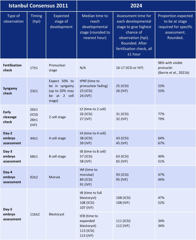
One major change is the move away from traditional days post-insemination, such as Day 1 and Day 2. The new unified benchmark uses hours post-insemination (hpi).
This change addresses differences among embryos labeled as being on the same day. It also provides a stronger basis for standardizing data across laboratories and culture conditions.
Based on extensive clinical research, the Consensus identifies three main factors that influence embryo development timing:
Impact of fertilization method on early development
Fertilization method affects the timing of the first cleavage. ICSI embryos reach the 2-cell stage (t2) 0.98 hours earlier than IVF embryos (26 hpi versus 27 hpi). However, blastocyst formation begins 1.157 hours later in ICSI embryos. A fully formed blastocyst is reached 1.510 hours later. This discrepancy indicates that early cleavage speed cannot fully represent subsequent developmental potential. Assessment should consider the embryo’s full kinetic profile. Relying on one time point may lead to incorrect judgments about viability.
Some ICSI embryos show rapid early cleavage but later arrest during blastocyst formation. Cell rearrangement or gene-expression regulation may contribute to this outcome.
Temporal characteristics of aneuploid embryos
Studies report significant delays in 10 morphokinetic parameters among aneuploid embryos. These delays extend from the 8-cell stage (t8) to the expanded blastocyst stage. Longer or shorter cell cycles may reflect several biological problems. These include abnormal DNA repair, impaired cell rearrangement, and failed cell-cycle checkpoints. This finding provides an important basis for the preliminary screening of euploid embryos using TLT. This helps laboratories narrow down the screening range before genetic testing.
Critical role of the culture environment
Environmental factors significantly affect embryo development speed. Embryos cultured at 20% oxygen develop more slowly than those cultured at 5% oxygen. The higher concentration is also associated with a lower implantation rate. A shift in medium pH toward alkalinity or a decrease in temperature slows embryo development. Culture-medium type and composition also influence embryo morphokinetics. Researchers should account for these differences when comparing results. Ovarian stimulation protocols may change early cleavage timing. Current evidence does not show an effect on overall embryo quality.
Laboratories must strictly control the stability of the culture environment, record the protocols used, and ensure data traceability.
Changes in Pronuclear Stage Assessment
The pronuclear stage is important for assessing fertilization. It also affects how later developmental potential is evaluated. New findings from TLT and PGT-A led to several adjustments. They cover assessment timing, pronuclear-number terminology, and clinical selection.
The Consensus also identifies indicators that currently lack a confirmed link with embryo quality. This clarification helps prevent overinterpretation:
1. Assessment Timing: 16–16.5 hpi as the Optimal Static Observation Window
The 2011 Consensus recommended pronuclear assessment at 17 ± 1 hpi. The new Consensus draws on a large TLT study of IVF and ICSI embryos. It identifies 16–16.5 hpi as the optimal window for static pronuclear counting.
During this window, visibility of two pronuclei (2PN) exceeds 98%. This timing therefore maximizes detection of normally fertilized zygotes.
After 17 hpi, some pronuclei may already have undergone pronuclear breakdown (PNBD). They may then be misclassified as unfertilized, potentially leading to the loss of viable embryos. Therefore, laboratories must strictly adhere to this pronuclear assessment time window to avoid timing-related inaccuracies.
2. Pronuclear Number: Updated Terminology and Clinical Value Judgment
“Pronuclei not observed” replaces “0PN”
Some embryos develop normally even when pronuclei are not seen during static observation. For these cases, the Consensus replaces 0PN with the term pronuclei not observed. Overall morphokinetic evidence does not confirm that “embryos without pronuclear formation can still develop.” On the contrary, it is highly likely that “0PN” embryos that progress to the first mitosis are actually 2PN, or in rare cases 1PN/multiple pronuclei. This underwent PNBD before pronuclei could be detected during static fertilization assessment.
PNBD may occur before the traditional assessment time. Static observation can therefore miss it. These embryos may still have normal developmental potential and live birth rates comparable with 2PN embryos. Such embryos should not be directly classified as “unfertilized.” In fact, embryos that exhibit faster morphokinetic characteristics during the fertilization stage generally have stronger developmental capabilities.
1PN pronuclei
In the past, 1PN embryos were often regarded as abnormal and discarded. However, PGT-A technology has rehabilitated their clinical value. Studies indicate that approximately 40%–50% of 1PN blastocysts are biparental diploids with a normal chromosome composition when tested by PGT-A. In terms of implantation rate, pregnancy rate, and live birth rate, 1PN blastocysts perform excellently, comparable to traditionally normally fertilized 2PN blastocysts.
Exploring their formation mechanism: in IVF cycles, some 1PN embryos result from asynchronous development of male and female pronuclei or their premature fusion. Although these embryos appear as “single pronuclei,” they have intact chromosome sets and the potential to develop into healthy fetuses. However, 1PN embryos are not without risks: in ICSI cycles, the blastocyst formation rate of 1PN embryos (17.4%) is significantly lower than that in IVF cycles (33.7%).
This indicates that the fertilization method has a significant impact on the subsequent development of 1PN embryos.
2.1PN pronuclei
2.1PN pronuclei—referring to zygotes with 2 normal pronuclei accompanied by 1 micronucleus—are extremely rare, accounting for less than 1% of embryos. Previously, due to technical limitations and their “abnormal” pronuclear composition, they were often directly discarded in clinical practice. However, with in-depth research, especially the accumulation of clinical cases in recent years, it has been confirmed that 2.1PN embryos can also develop into biparental diploid blastocysts and eventually result in the birth of healthy-appearing infants.
Relevant studies show that the developmental potential of 2.1PN embryos is influenced by multiple factors: 2.1PN zygotes from older patients (≥38 years old) have stronger blastocyst formation ability than those from younger patients, and the difference in developmental potential compared with 2PN zygotes narrows. When determining whether 2.1PN embryos are worth culturing, the early embryo cleavage pattern and Day 3 embryo quality are key predictive indicators—embryos with a normal cleavage pattern and high-quality grading have a higher chance of blastocyst formation.
3PN pronuclei
Most 3PN embryos result from digynic/diandric fertilization and carry an extremely high risk of aneuploidy. Clinical application cases are extremely rare, so routine use is not recommended for the time being. However, preclinical research is encouraged to explore their potential value (e.g., whether rare euploid cases exist).
3. Morphological Characteristics: Clarifying Dynamic Indicators “Insufficiently Proven to Be Relevant”
Many morphological characteristics of the pronuclear stage show dynamic changes, and current evidence is insufficient to confirm a strong association with embryo quality. Overreliance on these indicators during clinical assessment should be avoided:
Pronuclear size and position
The paternal pronucleus is usually larger than the maternal pronucleus, and the size difference gradually decreases as fertilization progresses. However, existing studies have conflicting conclusions on whether “pronuclear size difference affects outcomes,” so it cannot be confirmed as a reliable predictive indicator. Only “extremely rare eccentric positioning” (with an extremely low incidence) of pronuclei is associated with abnormal cleavage. Occasional positional differences observed in routine static observation are insufficient as a basis for excluding embryos.
Nucleolar precursor body (NPB) pattern
The process of NPBs from condensation and aggregation to dispersion is continuous and dynamic. There are differences in the aggregation kinetics of NPBs between paternal and maternal pronuclei, and even after aggregation is complete, NPBs may actively disperse a few hours before pronuclear breakdown. Current studies show that NPB patterns have no clear predictive value for implantation and live birth outcomes. Only studies that complexly calculate their movement speed have found weak correlations. This is insufficient to qualify NPB patterns as clinical assessment indicators.
Cytoplasmic halo
A cytoplasmic halo is a region with reduced cytoplasmic granularity in the pronuclear cortical area, visible in 82%–98% of pronuclei. It typically forms 2–4 hours after pronuclei appear and disappears 1 hour before pronuclear breakdown. Studies have confirmed that the absence of a cytoplasmic halo affects blastocyst formation efficiency but does not impact the implantation rate of blastocysts after transfer. Thus, it can be used for ranking embryos in Day 3 embryo transfers but cannot serve as a basis for excluding embryos.
If embryos are cultured to the blastocyst stage, the presence or absence of a cytoplasmic halo has no significant impact on outcomes.
In summary, the updates to the embryo development timeline and pronuclear stage assessment in the new Consensus provide more precise standardized guidelines for clinical practice. The development timeline uses hpi as the benchmark. It is recommended that each center establish a reasonable assessment time based on its own dataset.
Adjustments to the pronuclear stage are adapted to the application of time-lapse incubator systems. This reduces misjudgments. The interpretation of cleavage stage and blastocyst stage assessment key points will be covered in subsequent articles.